Endosonography is a high-tech ultrasound study that simultaneously combines the possibilities of endoscopic and ultrasound diagnosis of diseases of the gastrointestinal tract, pancreas, bile ducts and liver.
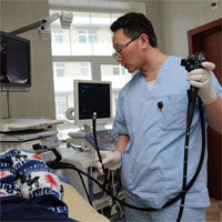
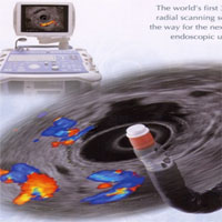
The study is carried out using a video endoscope, at the end of which there is a radially scanning ultrasonic sensor. The use of very high ultrasonic frequencies (5.0, 7.5, 12, and 20 MHz) in the device provides a high quality image with a resolving power of less than 1 mm that is not available to other methods of investigation such as conventional ultrasound, computer and magnetic resonance imaging, endoscopic cholangiopancreatography. At the same time, endosonography is not associated with the risk of radiological exposure to personnel and the patient, there is no risk of complications inherent in ERCP.
The advantages of endoscopic ultrasound before the traditional ultrasound examination through the front wall of the abdomen are that the ultrasonic sensor can be guided directly to the object under examination by the lumen of the digestive tube under visual control.
The wall of the digestive tube with ultrasound visualization is represented as alternating dark and light strips, each of which corresponds to the mucous, submucosal, muscular, adventitious layers with their interlayers. Thickening of certain layers, a violation of their regularity, clarity of boundaries and other changes allow us to determine the presence of a pathological focus and to assess its distribution deep into the wall and beyond. The use of endosonography in tumorous diseases of the abdominal cavity makes it possible to detect altered regional lymph nodes.
The depth of penetration of ultrasound into the tissues surrounding the digestive tube with a clear visualization is up to
The main indications for the use of endosonography:
- Diagnosis of volumetric formations of the pancreas, BDS, intracerebral tumors, as well as the stages of their spread. Identification of regional and distant metastases in lymph nodes
- Determination of the stage of the malignant process and the depth of the lesion with a small size of education.
- Identification of gallstones in the bile ducts without the use of ERCP.
- Diagnosis of the severity of changes in the parenchyma and ducts, pancreas for various types of chronic pancreatitis and its complications.
- Submucosal tumors of the upper gastrointestinal tract or suspected of their presence by endoscopic examination.


