Diagnostic Methods
Radiography
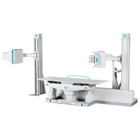
The radiography laboratory conducts research in the following directions:
- Lungs research;
- Research of bone structures (small and large joints, ribs, collarbones, sternum, skull, bones of the facial skull, pelvic bones, spine, including functional tests);
- Studies of ENT organs (accessory sinuses, nasal and oropharynx);
- Contrast study of the urinary system (excretory urography).
Mammography
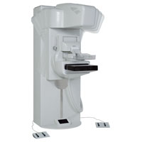
Radiographic mammography can detect pathological changes in the breast and determine their nature. Currently, mammography is one of the main diagnostic techniques for fibrocystic disease, fibroadenoma and malignant neoplasms of the breast. Our diagnostic center uses the unique full-scale digital mammography system of the latest generation with the possibility of three-dimensional tomosynthesis.
Most of the research is done in a planned manner, in the direction of the doctor, for the final confirmation of the diagnosis. There are studies that are performed with contrast medications.
The success of research in the department of radiation diagnosis is directly dependent on the qualitative preparation of patients for them. The nature of preparation depends entirely on the type of equipment used, on the chosen technique of research and the organ being examined.
CT scan
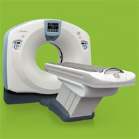
Computer tomography is a modern
In this modification of the CT scanner, an oncopy program is installed that will allow not only visualizing the formations from 2 to 12 mm, but also differentiating from other formations and assessing changes in dynamics. There is also a program for diagnosing lung diseases, to assess the severity of emphysema.
In the department of radiation diagnosis the following CT studies are carried out:
- Lungs, mediastinum;
- Brain;
- Organs of the neck, face, paranasal sinuses, larynx;
- Abdominal cavity organs;
- Urogenital system;
- Osteoarticular system (joints, bones, spine).
- Studies of abdominal organs with contrast;
- Study of organs of the genitourinary system with contrasting;
- Angiography of vessels: large vessels of the thoracic and abdominal cavities, vessels of the brain, brachiocephalic vessels, vessels of the upper and lower extremities.
Preparation for X-ray studies
-
X-ray examinations of the thoracic cavity, heart with contrasted esophagus, larynx, paranasal sinuses, bones of the skull, limbs, cervical and thoracic spine are carried out without special preparation of the patient. - In the study of the lumbosacral spine, pelvic bones, bladder, uterus, a review enema requires a cleansing enema the night before. Patients come to study after a light breakfast.
- Intravenous urography is performed on an empty stomach. It is mandatory that the attending physician mark in the medical guideline with the formulation: “There are no contraindications to intravascular radiopaque studies. The patient’s consent to the study was received.” The night before, a cleansing enema or other preparation of the intestine is carried out.
- Mammography is performed on the
8-10 day from the beginning of the menstrual cycle. - Metrosalpingography is performed on an empty stomach, with a prepared intestine.
Preparing for X-ray computed tomography
- Standard (non-contrast) CT examinations of the bones of the skull, brain, paranasal sinuses, temporal bones, neck, thyroid, larynx, thoracic cavity, mediastinum, spine, scapula, large joints, limbs are carried out without preliminary preparation of patients.
- Contrastless CT examination of the abdominal cavity (liver, spleen, pancreas, kidneys and adrenal glands) is performed on an empty stomach.
- CT examinations of various organs and systems using contrast intravenous enhancement are performed on an empty stomach on the recommendation of a radiologist and on the appointment of a doctor in charge, after a careful study of the patient’s allergic history, no contraindications for intravenous radiocontrast agents. On the eve of the study (the previous day), the patient should drink
1-2 liters of water (liquid) additionally. - The condition for conducting CT cardioangiography (CT CAG) is:
- The heart rate at rest should not exceed 65 beats / min;
- Absence of interruptions in the work of the heart (extrasystoles and other rhythm disturbances);
- The ability to hold your breath during the study for 25 seconds.
Attention! It should be remembered that CT CAG does not replace invasive CAG, but is a screening diagnostic method.
Contact Information
Address: Moscow, Gagarinsky lane, 37 (metro station Smolenskaya of the Arbatsko-Pokrovskaya line or metro station Kropotkinskaya)
Phone:


