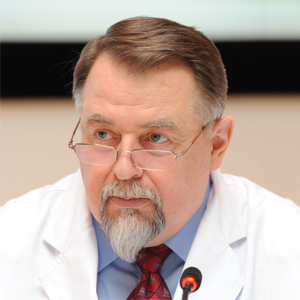
Chief of the department: Kanshina Darya Sergeevna, M.D., Ph.D., neurologist of the highest category, a physician of functional diagnostics.
What do we treat
Neuromuscular diseases
Methods of research:
- ENMG (EMG) is a set of techniques that allows you to verify the level and severity of lesions of the neuromuscular system. Currently, there are more than 20 modalities of ENMG (EMG), the most common are: stimulation, needle, surface EMG.
- Diagnostic magnetic stimulation — the effect of pulsed magnetic field on various parts of the cortex of the brain, spinal cord, peripheral nerves — is widely used for spinal trauma, myelopathy (spinal cord injury of vascular genesis), demyelinating processes (multiple sclerosis, etc.), period of silence, diagnosis of Parkinson’s disease.
- Ultrasound examination of peripheral nerves and muscles is a non-invasive method for studying the state of the neuromuscular apparatus, which allows revealing the posttraumatic, autoimmune nature of nerve damage, neoplasms, and the severity of the nerve trunk edema in compression tunnel neuropathies.
Epilepsy
Methods of research:
- Electroencephalography (EEG) with functional tests is a routine EEG that is performed according to international standards, lasting
15-20 minutes with registration of bioelectric activity at rest and during hyperventilation and photostimulation. It is a screening technique, which allows to reveal anomalies of activity of neurons of the brain. However, this record does not always allow verification of paroxysmal (including epileptiform) activity, which requires a more in-depth examination (usually in patients with suspected epilepsy). - Video-EEG monitoring is a multi-hour recording of the EEG for the purpose of recording an epileptiform attack. EEG is evaluated in combination with clinical manifestations of the disease.
Indications for video EEG monitoring:
- Diagnosis of the form of epilepsy.
- Differential diagnosis of paroxysmal conditions — fainting, loss of consciousness, fading for a few seconds, sudden dizziness, unmotivated behavior, a fragmentary loss of memory.
- Survey of candidates for surgical treatment of epilepsy — evaluation of inter-attack activity, registration of an attack to determine the source of the epileptogenic zone, analysis of the seizure.
- Diagnosis of psychogenic non-epileptic paroxysms.
- Sleep disturbances (nighttime fears, nightmares, sleepwalking, sleeptalking).
Complex diagnosis in the pelvic nerves pathology
Diagnosis of pelvic nerves pathology includes a study of the conduct on the pudendal nerves with the use of electroneuromyography, magnetic stimulation, evoked potentials. The main indications for the study are: a dysfunction of the pelvic organs, neuropathic pain in the perineum, erectile dysfunction. The obtained results of the study allows to determine the further tactics of treatment (conservative therapy or surgical intervention).
Multimodal examination of the central and peripheral nervous system in various conditions and diseases (demyelinating diseases, Alzheimer’s disease, etc.)
The evoked potentials (EP) is a method of recording the responses of various brain structures to external stimuli, auditory, visual and somatosensory, evaluation of the ascending central nervous system.
The evoked potentials are used for a wide range of lesions of the central nervous system for objectifying the lesion, determining its level and character.
- Visual evoked potentials: registration of visual cortex responses to stimulation with a reversal pattern or light flash, the visual pathways from the retina to the occipital cortex are examined. Allow to diagnose lesions of the optic nerve (retrobulbar neuritis, ischemic neuropathy), retrochiasmal lesions — the visual tract, are widely used in the diagnosis of multiple sclerosis.
- Auditory evoked potentials: recording of impulses along the peripheral and central sections of the auditory analyzer. Used for differential diagnostics of central and peripheral lesions of the acoustic system, are extremely useful in diagnosing lesions of the cerebellar angle, are highly sensitive to multiple sclerosis, often in the absence of clinical symptomatology of the trunk.
- Somatosensory EP from the hands and feet: a study of the sensory pathways of the central nervous system, the responses of the spinal cord and brain to electrical stimulation of the peripheral nerves. Evaluation of demyelinating, degenerative and vascular lesions of the central nervous system, can be used in the diagnosis of plexopathy and radiculopathy, as a confirmatory test for diabetic polyneuropathy, etc.
- Cognitive EP P300 and MMN — this kind of evoked potentials is an indicator of bioelectric processes associated with the mechanisms of perception of external information and its processing. The essence of the method lies in the analysis of endogenous events occurring in the brain, associated with the recognition and memorization of the stimulus. Assessment of cognitive deficits (chronic cerebral ischemia with cognitive disorders, dementia, Alzheimer’s disease, etc.)
Intraoperative neurophysiological monitoring
One of the newest methods, which is a complex of studies: electrocorticography, evoked potentials and myography, used during surgical neurosurgical interventions on the brain and spinal cord, with the installation of stabilizing systems, with surgical interventions for the defeat of peripheral nerves. It allows to evaluate the conductive capacity of the nervous system, its slightest changes in the course of surgical intervention, thereby reducing the risk of developing a neurologic deficit in the postoperative period, improving the patient’s quality of life. According to the order of the Ministry of Health is an obligatory technique for high-tech neurosurgical care for the population in a number of neurological pathologies.
Also on the basis of the Pediatrical Consultative and Diagnostic Center of the National Pirogov Medical and Surgical Center the following types of research are conducted for children from 1 year: stimulation and needle electroneuromyography, diagnostic magnetic stimulation, pelvic examination, video EEG monitoring during daytime sleep (in the ward of a day hospital).
Contact Information
Address: 105203, Moscow, Nizhnyaya Pervomayskaya str., 70
Contact phone number:
Fax:
How to reach us by using public transport
“Pervomaiskaya” metro station (last carriage of the train out of the city centre). From “Pervomaiskaya” metro station by any tram or trolley bus go to the stop “15th Parkovaya Street”. Go along the 15th Park Street to the intersection with the Nizhnyaya Pervomaiskaya Street, turn left and walk about a hundred meters to the entrance of the Pirogov National Medical and Surgical Center.


